Molecular imaging is an important tool during both the preclinical and clinical stages of therapeutic development. Agents that specifically localize to the target tissue and demonstrate little secondary accumulation in nontarget tissues can be selected by employing molecular imaging to maximize therapeutic efficacy and minimize harmful side effects. Molecular imaging allows for the visualization of cellular and subcellular processes at both the molecular and anatomical levels. It can provide not only the visualization of biological and pathophysiological processes, but also characterization and quantification of these processes. The techniques used in molecular imaging typically rely on exploiting disease-specific molecular probes and intrinsic tissue characteristics. Currently, molecular imaging is used for diagnostic/prognostic purposes and to measure therapeutic efficacy in preclinical models and patient populations in the clinic.
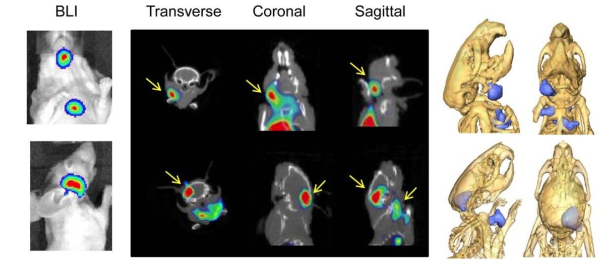
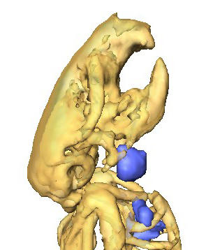
Our lab specializes in optical and nuclear imaging. Fluorescence imaging uses near infrared dye-conjugated antibodies and is an excellent laboratory tool. Positron emission tomography (PET) nuclear imaging uses radiolabeled antibodies with tracers such as zirconium-89. This imaging modality is the most clinically relevant.